
GLP-1 pharmacokinetic research measures absorption, distribution, metabolism, and excretion using bioanalytical methods such as LC-MS/MS for quantifying plasma levels, with HPLC used for purity and degradation checks. Native GLP-1 clears within minutes because DPP-4 degrades it, while acylated analogs extend half-life to about a week, so sampling schedules must match the analog.
Key points
- GLP-1 pharmacokinetic research depends on LC-MS/MS or HPLC to track half-life, Cmax, and Tmax accurately.
- Acylated and dual-agonist GLP-1 analogs need different sampling windows than native peptide sequences.
- Reconstitution inconsistency is the most common source of PK data variability in academic and CRO labs.
- GLP-123 supplies research peptides with certificates of analysis so identity is confirmed before a study starts.
GLP-1 pharmacokinetic research maps how a peptide analog moves through absorption, distribution, metabolism, and excretion (ADME), and the method you pick determines whether your half-life, Cmax, and Tmax numbers hold up under peer review. For laboratory and contract research teams, that means choosing a bioanalytical method, a sampling schedule, and a reconstitution protocol before the first dose ever leaves the vial.
- GLP-1 pharmacokinetic research depends on LC-MS/MS or HPLC to track half-life, Cmax, and Tmax accurately.
- Acylated and dual-agonist GLP-1 analogs need different sampling windows than native peptide sequences.
- Reconstitution inconsistency is the most common source of PK data variability in academic and CRO labs.
- GLP-123 supplies research peptides with certificates of analysis so identity is confirmed before a study starts.
Why GLP-1 pharmacokinetic research matters for lab and CRO teams
GLP-1 receptor agonists span a wide range of elimination profiles. Native GLP-1 clears from circulation within minutes because dipeptidyl peptidase-4 (DPP-4) degrades it almost immediately, while structurally modified analogs that use fatty-acid acylation or albumin binding extend half-life to roughly a week, a pharmacology fact documented across published prescribing information for once-weekly agents. That spread means a PK protocol built for one analog class rarely transfers cleanly to another.
For academic and CRO labs working under fixed budgets and shared instrument time, the cost of a mismatched protocol isn't just wasted reagent. A sampling schedule that misses the elimination phase for a long-acting analog produces a half-life estimate that's simply wrong, and a wrong number that gets published or handed to a client is worse than no number at all. The translational GLP-1 research methods used to bridge in vitro binding data to in vivo exposure data exist specifically to catch this kind of mismatch before it reaches a report.
The GLP-1 pharmacokinetic research workflow, step by step
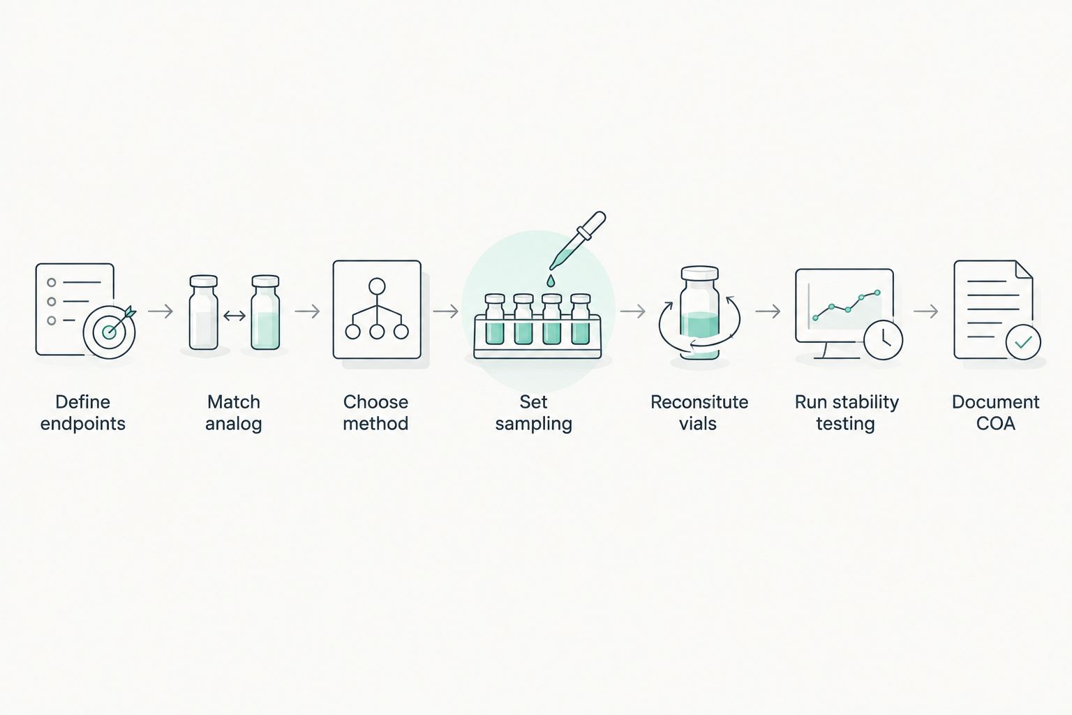
Define your pharmacokinetic endpoints first
Every downstream decision, from analog selection to sampling density, follows from the endpoints you're actually trying to measure. Pick these before you touch a pipette.
- Cmax and Tmax, the peak concentration and time to reach it
- Area under the curve (AUC), for total exposure
- Terminal half-life (t1/2), for dosing interval decisions
- Clearance and volume of distribution (Vd), for compartmental modeling
- Bioavailability (F%), if comparing routes of administration
Match the analog and formulation to the study question
Not every GLP-1 analog behaves the same way in a PK model, and the study question should dictate which one you use.
- Native GLP-1 sequence for baseline degradation kinetics
- Acylated, albumin-binding analogs for extended half-life characterization
- Dual or triple receptor agonists for cross-reactivity and receptor desensitization studies
- Lyophilized stock for long-term storage stability arms
- Single-lot vs multi-lot comparison when testing for batch-to-batch variability
Choose the bioanalytical method before you dose the first sample
The assay you pick has to match the sensitivity and specificity the endpoint demands, and switching methods mid-study invalidates your calibration curve.
- LC-MS/MS for quantitative plasma-level tracking, the standard for peptide PK work
- HPLC for purity and degradation monitoring rather than plasma quantification
- Immunoassay-based methods (ELISA) as a lower-cost screening proxy for receptor binding
- Radiolabeled tracer studies for tissue distribution mapping in animal models
- Method validation against a certified reference standard before any real samples run
Bioanalytical methods built specifically for GLP-1 peptide classes cover the validation criteria (linearity, lower limit of quantification, matrix effect) that separate a defensible assay from a guess.
Set the sampling schedule around the analog's expected half-life
A sampling schedule built for a fast-clearing peptide will completely miss the terminal elimination phase of a long-acting analog.
- Dense early time points to catch Cmax and Tmax accurately
- Extended terminal sampling matched to the analog's expected elimination phase
- A matrix effects check comparing plasma vs. serum before finalizing collection tubes
- Fixed time-to-assay windows so degradation during storage doesn't skew early vs. late samples
Reconstitute and handle every vial the same way
This is where most PK data variability actually originates, and it's fixable with discipline rather than new equipment. Reconstitution volume changes effective concentration, and an inconsistent volume across dosing groups introduces error before the animal or cell model even sees the compound.
- Use a fixed bacteriostatic water volume on every reconstitution, no exceptions
- Record the reconstitution date and storage condition against every vial
- Discard any vial showing turbidity or visible particulate
- Log lot numbers against dosing groups so a bad lot is traceable
- Run a pre-dose HPLC check on actual concentration when precision matters
This is the point where sourcing quality starts to matter as much as protocol design. GLP-123 ships research peptides with a certificate of analysis per lot, which shortens the identity-verification step this section describes rather than replacing it.
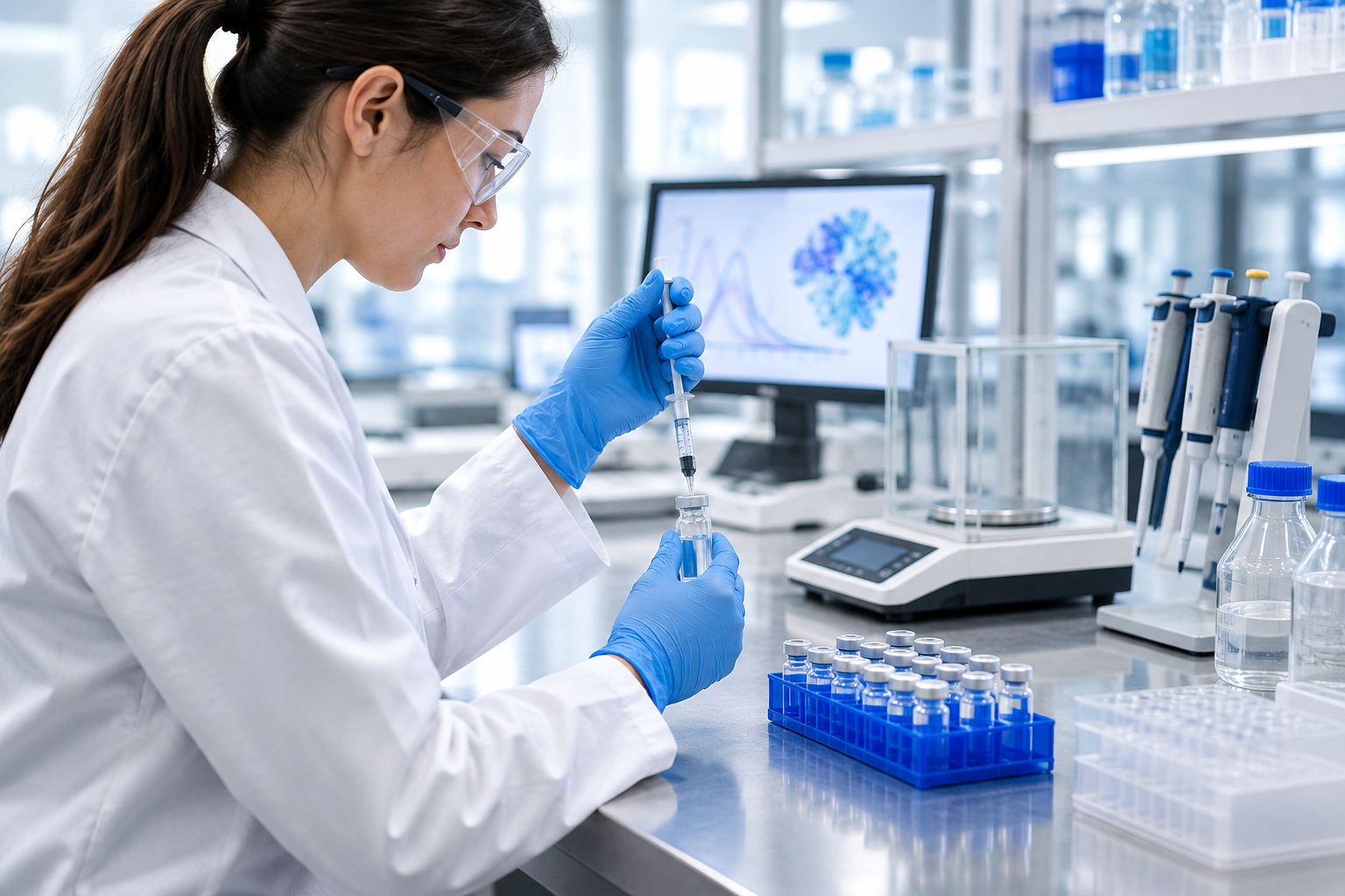
For the mechanics of the reconstitution step itself, the guide on reconstituting GLP-1 research peptides walks through syringe technique and vial handling in more detail.
Run stability and degradation testing alongside the PK study
A PK dataset is only as good as the compound's stability during the sampling window. Build stability checks into the same timeline rather than treating them as a separate project.
- Forced degradation testing under heat and light stress
- Freeze-thaw cycle tracking for any vial reused across multiple dosing days
- Real-time vs. accelerated stability arms run in parallel
- Endotoxin screening before any in vivo dosing
Document everything with a certificate of analysis and a raw data trail
A report without raw data behind it doesn't survive review. This step is administrative, not optional.
- Retain the certificate of analysis (COA) for every lot used in the study
- Log every assay run against the instrument's calibration ID
- Keep raw chromatograms, not just summary tables
- Cross-reference purity data against your dosing calculations before finalizing results
Comparing PK research method options
| Option | Best for | Key limitation |
|---|---|---|
| In-house LC-MS/MS assay | Teams needing quantitative plasma-concentration data | Requires calibrated instrumentation and a trained analyst on staff |
| Outsourced CRO bioanalytical service | Labs without in-house LC-MS access | Turnaround depends on the CRO's queue, less real-time control |
| HPLC purity and degradation tracking | Stability arms and batch-release testing | Confirms identity and purity, doesn't quantify plasma exposure |
| Reference standard plus COA verification | Confirming peptide identity before dosing | A pre-check step, not a substitute for full PK modeling |
In-house LC-MS/MS wins on data control for teams that already own the instrument; outsourcing to a CRO wins for labs running occasional studies without justifying a standalone bioanalytical setup.
Common mistakes academic and CRO labs make in GLP-1 PK research
- Skipping the pre-dose purity check. Assuming a vial matches its label without a confirmatory assay turns every downstream number into a guess.
- Changing bacteriostatic water volume between dosing groups. This shifts effective concentration and directly confounds Cmax comparisons across the same study.
- Using one terminal sampling point for peptides with different half-lives. A schedule built for a fast-clearing analog misses the elimination phase entirely for a long-acting one.
- Not logging temperature excursions between reconstitution and dosing. A few hours at room temperature can degrade a peptide enough to shift the exposure curve.
- Comparing PK data across studies run on different LC-MS calibration curves. Without a shared reference standard, the comparison isn't valid no matter how clean either dataset looks alone.
FAQ
What is GLP-1 pharmacokinetic research?
GLP-1 pharmacokinetic research studies how a GLP-1 peptide analog is absorbed, distributed, metabolized, and excreted, measuring endpoints like half-life, Cmax, Tmax, and AUC. It’s the standard method labs use to characterize exposure before scaling a study.
Which assay is best for GLP-1 PK studies, LC-MS or HPLC?
LC-MS/MS is the standard for quantifying plasma concentration in GLP-1 pharmacokinetic research. HPLC is better suited to purity and degradation tracking rather than plasma-level quantification.
Why do GLP-1 analogs have such different half-lives?
Structural modifications like fatty-acid acylation and albumin binding extend circulation time by resisting DPP-4 degradation. Native GLP-1 clears in minutes; modified once-weekly analogs can circulate for close to a week, per published prescribing data.
How does reconstitution affect PK study accuracy?
Reconstitution volume sets the effective concentration of the peptide solution. An inconsistent volume across dosing groups introduces concentration error before the sample is even dosed, which is the most common source of PK data variability in lab settings.
What should a certificate of analysis include for a GLP-1 research peptide?
A COA should show identity confirmation, purity percentage, and lot-specific testing data tied to the exact vial used in the study. Retaining this per lot keeps the study’s raw data trail defensible during review.
Can PK data from different studies be compared directly?
Only if both studies ran on the same or a cross-validated LC-MS calibration curve and used the same reference standard. Without that, differences in the data may reflect assay variation rather than a real biological difference.
Do animal models or cell-based assays give better PK data?
Cell-based assays are faster and cheaper for early screening of binding and receptor activity, but only animal models capture full ADME behavior including distribution and excretion. Most protocols use both in sequence.
One last thing
The single variable that wrecks more GLP-1 pharmacokinetic research datasets than any instrument choice is reconstitution consistency. Labs troubleshoot LC-MS calibration curves for weeks looking for a source of variability that turns out to be a bacteriostatic water volume that drifted between technicians. Fix that one step and most PK data noise disappears before you touch the assay.
Related guides
Originally published on glp-123.com. This library mirrors the guide for easier reading. Products mentioned are for laboratory research only.
View original


