
Develop GLP-1 receptor assays by confirming peptide identity and purity with LC-MS/HPLC first, then measuring receptor affinity by binding, functional signaling by cAMP, and biased signaling by β-arrestin recruitment when relevant. Stable human GLP-1R-expressing CHO-K1 or HEK293 cells are standard for binding and cAMP assays.
Key points
- GLP-1 assay development runs four stages in order: binding, cAMP signaling, β-arrestin recruitment, then purity confirmation by LC-MS/HPLC.
- Radioligand binding assays measure receptor affinity (Ki/Kd); cAMP assays confirm the receptor actually signals.
- β-arrestin recruitment assays separate biased agonists from full agonists — critical for comparing analog candidates.
- Reagent purity below the 95% research-grade threshold is the most common cause of inconsistent EC50 curves.
Developing a GLP-1 receptor assay in 2026 means choosing among four core formats — radioligand binding, cAMP accumulation, β-arrestin recruitment, and LC-MS/HPLC purity confirmation — and running them in that order so binding and functional data aren't corrupted by degraded peptide. The step most labs skip is confirming reagent purity before the assay, not after: a peptide that's 85% pure instead of the research-grade ≥95% purity by HPLC standard will shift your EC50 curve and make a perfectly good receptor construct look broken.
- GLP-1 assay development runs four stages in order: binding, cAMP signaling, β-arrestin recruitment, then purity confirmation by LC-MS/HPLC.
- Radioligand binding assays measure receptor affinity (Ki/Kd); cAMP assays confirm the receptor actually signals.
- β-arrestin recruitment assays separate biased agonists from full agonists — critical for comparing analog candidates.
- Reagent purity below the 95% research-grade threshold is the most common cause of inconsistent EC50 curves.
- CHO-K1 and INS-1 lines overexpressing GLP-1R are the standard cell models for functional readouts in 2026.
Why this matters
A GLP-1 receptor assay is only as good as the reagent behind it. Labs running GLP-1 receptor structure research know the receptor is a Class B GPCR with a large extracellular domain that binds the peptide N-terminus first, then folds the C-terminal helix into the transmembrane bundle — a two-step binding mechanism that determines which assay format actually detects your analog.
Get the assay wrong and you don't just lose a week. You publish or internally report a Ki or EC50 that doesn't reproduce, and every downstream comparison against a reference agonist becomes noise. This is why assay development for GLP-1 receptor work in 2026 starts with reagent qualification, not with plating cells.
How to develop GLP-1 receptor assays
The development sequence below reflects how contract research labs and university groups structure GLP-1 assay development work in 2026.
| Assay Type | What It Measures | Best For | Typical Readout |
|---|---|---|---|
| Radioligand binding | Receptor affinity (Ki, Kd) | Screening analog binding potency | Bound vs. free counts |
| cAMP accumulation | Downstream Gs signaling | Confirming functional agonism | Cyclic AMP concentration (HTRF/ELISA) |
| β-arrestin recruitment | Biased signaling, receptor desensitization | Comparing biased agonist profiles | BRET or PathHunter signal |
| LC-MS/HPLC | Peptide purity, identity, degradation | Confirming reagent quality before assay use | Retention time, mass fingerprint |
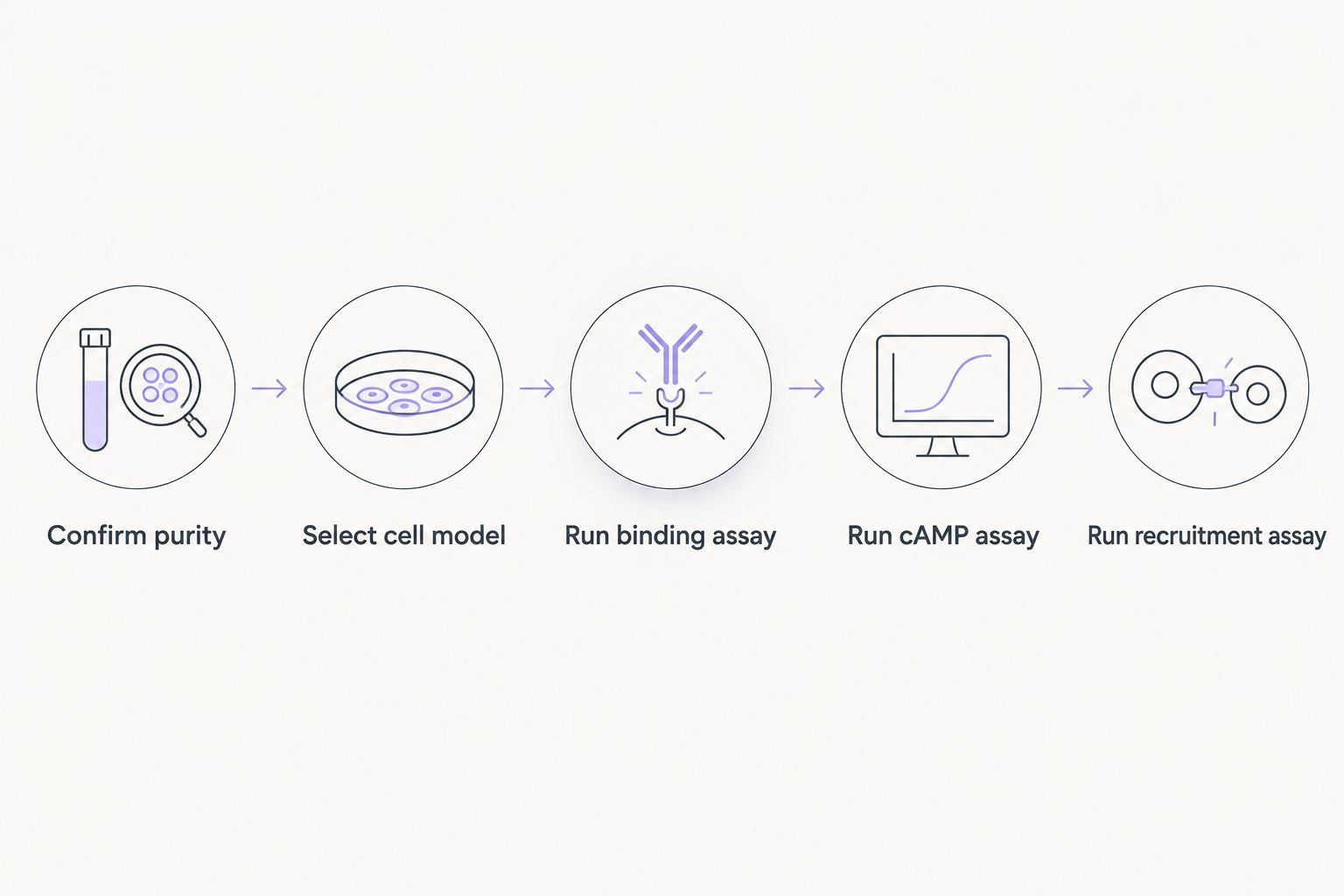
Steps, in order:
- Confirm reagent identity and purity by LC-MS/HPLC before any cell work — a peptide below 95% purity invalidates everything downstream.
- Select the cell model. CHO-K1 or HEK293 lines stably transfected with human GLP-1R are standard for binding and cAMP work; INS-1 832/13 cells are used when insulin-secretion coupling matters.
- Run the binding assay at 37°C, pH 7.4, using a radiolabeled or fluorescent tracer competed against unlabeled peptide to establish Ki.
- Run the cAMP functional assay in the same cell line to confirm the binding event produces a signaling response, not just occupancy.
- Run a β-arrestin recruitment assay if the goal is comparing biased agonists — G-protein and β-arrestin pathways diverge for several GLP-1 analogs in development.
- Document the certificate of analysis for every peptide lot used, since batch-to-batch purity drift is the single most common source of assay variance.
Radioligand Binding Assay: Confirming Receptor Affinity
Binding assays answer one question: does the peptide physically occupy the receptor, and how tightly? Competition binding against a labeled reference agonist gives you Ki, the standard metric labs report when comparing analog affinity against native GLP-1(7-36).
The main failure mode isn't the assay chemistry — it's the cell line. Endogenous GLP-1R expression in most immortalized lines is too low to get a usable signal, which is why overexpression constructs matter. Work built on GLP-1 receptor cell line models consistently shows CHO-K1 and HEK293 stable lines outperform transient transfection for binding reproducibility across replicate plates.
Verdict: use a stable overexpression line for binding work — transient transfection introduces too much well-to-well variance to trust a Ki comparison.
cAMP Accumulation Assay: Confirming Functional Signaling
Binding tells you the peptide sits on the receptor. cAMP accumulation tells you it actually turns the receptor on. GLP-1R couples primarily to Gs, so agonist binding should drive a measurable rise in intracellular cAMP, detected by HTRF or competitive ELISA formats.
This is the assay that separates a true agonist from a peptide that binds but doesn't activate — a distinction that matters when screening novel analogs or degraded lots that retain some binding affinity but have lost functional potency. Skip this step and you risk reporting binding data on a peptide that no longer signals.
β-arrestin Recruitment Assay: Detecting Biased Signaling
Some GLP-1 analogs activate G-protein signaling and β-arrestin recruitment to different degrees — a property called biased agonism, and it's a growing focus of comparative analog research in 2026. β-arrestin recruitment also drives receptor internalization and desensitization, which affects how long a signaling response lasts in cell-based systems.
BRET-based and enzyme-complementation assays (PathHunter-style) are the standard formats here. If your research question involves comparing two analogs that look identical in binding and cAMP data but behave differently in prolonged exposure studies, the divergence usually shows up in recruitment kinetics — a pattern documented in ongoing GLP-1 biased agonism research.
Verdict: run recruitment assays whenever the research question is analog comparison, not just confirming a single peptide works.
LC-MS/HPLC: Confirming Purity and Identity Before You Trust Any Result
Every assay above assumes the peptide going into the well is what the label says it is. LC-MS confirms molecular identity by mass; HPLC confirms purity by retention time and peak area. A peptide lot at 90% purity carries 10% of its mass as truncated fragments, oxidized variants, or synthesis byproducts — any of which can bind the receptor weakly and muddy a Ki curve without ever showing up as an obvious problem in the raw data.
This is the step research labs skip most often, usually because it feels like overhead rather than science. It isn't. A certificate of analysis with purity ≥95% and a matching mass spec fingerprint is the minimum bar before a GLP-1 peptide lot goes into any receptor assay.
Why GLP-1 Assay Sensitivity Varies
Assay-to-assay variance in GLP-1 receptor work traces back to a small number of recurring factors:
- Cell passage number — receptor expression drifts downward in overexpression lines after extended passaging.
- Peptide purity and storage — lyophilized peptides degrade faster once reconstituted and left at room temperature.
- Buffer pH and temperature — binding kinetics for Class B GPCRs are sensitive to deviations from physiological pH 7.4 and 37°C incubation.
- Tracer specific activity — radioligand or fluorescent tracer batches vary in labeling efficiency, shifting signal-to-noise.
- Reconstitution technique — inconsistent mixing or vial handling introduces peptide aggregation that reduces effective concentration.
- Reference standard lot — comparing new data against an old reference standard lot without re-validating introduces drift that looks like a real effect.
What cell line is best for GLP-1 receptor assays?
CHO-K1 and HEK293 lines stably transfected with human GLP-1R are the standard choice for binding and cAMP assays in 2026, because stable expression gives consistent receptor density across replicates. INS-1 832/13 cells are preferred when the research question involves insulin secretion coupling rather than receptor pharmacology alone.
Is a radioligand assay or a fluorescent binding assay better?
Radioligand binding remains the gold-standard reference method for Ki determination, but fluorescent/BRET-based binding assays are increasingly used in 2026 labs to avoid radioactive waste handling. Both formats produce comparable Ki values when tracer specific activity is properly characterized — the choice usually comes down to lab infrastructure, not sensitivity.
How do you know if a GLP-1 peptide lot is degraded before running an assay?
A degraded lot shows a shifted or split peak on HPLC and a mass discrepancy on LC-MS relative to the expected molecular weight. Purity below the 95% research-grade threshold is the practical cutoff most labs use to reject a lot before it reaches the assay bench.
FAQ
What is the best assay for measuring GLP-1 receptor binding affinity?
Radioligand competition binding is the standard method for measuring GLP-1 receptor binding affinity (Ki) in 2026. Fluorescent and BRET-based alternatives produce comparable data without radioactive waste handling.
How much peptide purity is needed for GLP-1 receptor assay development?
Research-grade purity of 95% or higher by HPLC is the standard threshold before a GLP-1 peptide lot is used in binding or functional assays. Below that, degradation byproducts distort Ki and EC50 values.
What cell line is used for GLP-1 receptor functional assays?
CHO-K1 and HEK293 cells stably overexpressing human GLP-1R are the standard functional assay models in 2026. INS-1 832/13 cells are used specifically for insulin-secretion coupling studies.
Is cAMP accumulation or β-arrestin recruitment better for comparing GLP-1 analogs?
cAMP accumulation confirms basic functional agonism, while β-arrestin recruitment reveals biased signaling differences between analogs. Comparative analog research typically runs both assays together.
Why does GLP-1 receptor assay data fail to reproduce between labs?
Reproducibility failures most often trace to peptide purity, cell passage number, or reference standard drift, not the assay chemistry itself. Confirming purity by LC-MS/HPLC before each run eliminates the most common source of variance.
What temperature and pH should GLP-1 binding assays run at?
GLP-1 receptor binding assays run at 37°C and pH 7.4 to match physiological conditions. Deviations from either shift binding kinetics enough to distort Ki comparisons across experiments.
Do you need a certificate of analysis for research peptides used in GLP-1 assays?
Yes — a certificate of analysis confirming purity and identity by mass spec is the minimum documentation before a peptide lot goes into a GLP-1 receptor assay. Without it, a failed assay can’t be traced back to reagent quality.
One last thing
The fastest way to shorten GLP-1 assay development timelines in 2026 isn't a faster plate reader — it's rejecting peptide lots at the purity-check stage instead of discovering the problem after a week of binding runs that won't reproduce. Labs that build LC-MS confirmation into their intake process, rather than treating it as optional QC, cut repeat-assay cycles the most.
Related guides
Originally published on glp-123.com. This library mirrors the guide for easier reading. Products mentioned are for laboratory research only.
View original


