
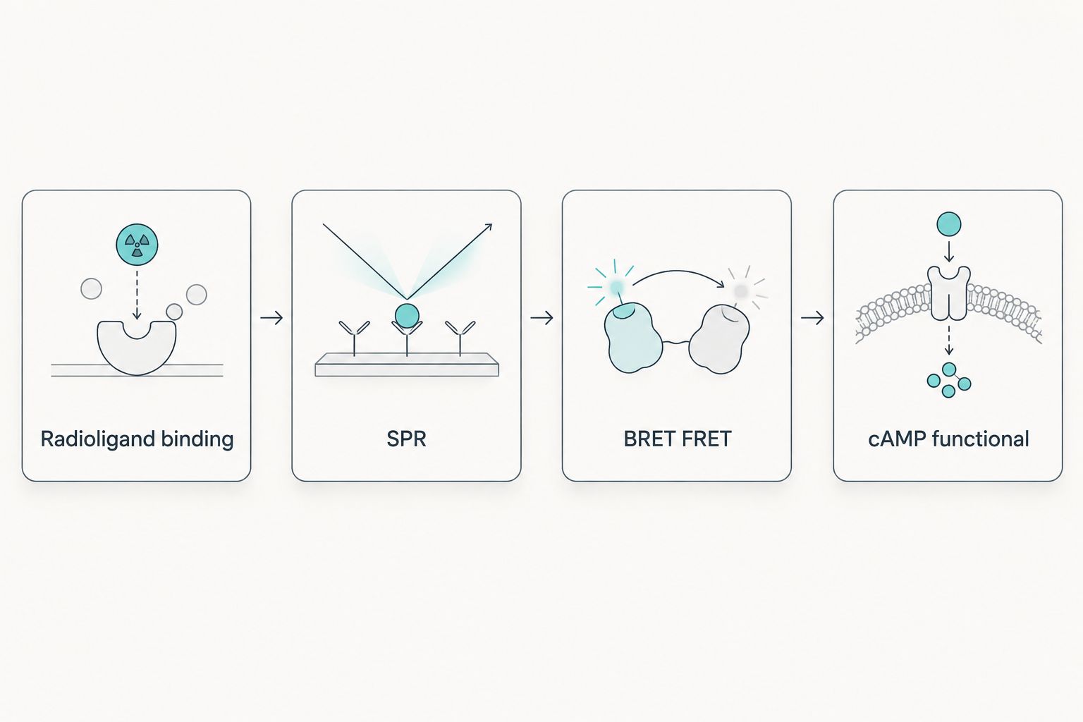
Radioligand competition binding in a 96-well format remains the reference method for generating comparable Kd and IC50 values. SPR provides label-free association and dissociation kinetics, and BRET/FRET suits higher-throughput live-cell screening. Binding alone does not predict signaling, so pair it with a functional cAMP readout.
Key points
- Radioligand competition binding in a 96-well format is still the reference method for a glp-1 receptor binding assay in 2026.
- SPR gives real-time association and dissociation kinetics without a label, but demands more purified peptide.
- Binding affinity alone doesn’t predict receptor signaling — pair it with a cAMP or biased-agonism readout.
- Cell line consistency, peptide purity, and buffer conditions drive most of the lab-to-lab variability researchers report.
A GLP-1 receptor binding assay measures how tightly a labeled or unlabeled ligand occupies the GLP-1 receptor (GLP-1R) on a target cell line, using one of three core formats: radioligand competition binding, surface plasmon resonance (SPR), or a fluorescence-based BRET/FRET readout. Radioligand competition binding, run in a 96-well plate with a radiolabeled or fluorescently tagged GLP-1 tracer incubated at 37°C for 60–90 minutes, remains the reference method for generating Kd and IC50 values in 2026 research protocols. The detail most protocols skip: binding affinity alone says nothing about downstream signaling, so a compound can look identical on a binding curve and behave completely differently in a functional cAMP assay.
- Radioligand competition binding in a 96-well format is still the reference method for a glp-1 receptor binding assay in 2026.
- SPR gives real-time association and dissociation kinetics without a label, but demands more purified peptide.
- Binding affinity alone doesn’t predict receptor signaling — pair it with a cAMP or biased-agonism readout.
- Cell line consistency, peptide purity, and buffer conditions drive most of the lab-to-lab variability researchers report.
Why this matters
Labs comparing published Kd or IC50 values against their own data run into a wall fast: assay format, cell line background, and tracer choice all shift the number, even when the underlying receptor pharmacology hasn't changed. A GLP-1 receptor assay development guide matters before you touch a plate, because the format you pick locks in what kind of data you can generate — occupancy, kinetics, or downstream signaling, not all three at once.
Getting the assay choice wrong wastes tracer, wastes peptide, and produces numbers that don't replicate across runs. That's the practical reason binding assay methodology gets more scrutiny in peer review than almost any other step in GLP-1 receptor pharmacology work in 2026.
How to study GLP-1 receptor binding in the lab
Four assay formats cover nearly all GLP-1 receptor binding work published today. Each answers a different question, and none of them substitutes for the others.
| Method | What it measures | Typical format | Best for |
|---|---|---|---|
| Radioligand competition binding | Displacement of a labeled tracer | 96-well plate, filtration or scintillation counting | Kd/IC50 generation, cross-study comparison |
| Surface plasmon resonance (SPR) | Real-time association/dissociation kinetics | Label-free, sensor chip | ka/kd rate constants, structure-activity work |
| BRET/FRET binding assay | Proximity-based fluorescence or luminescence signal | Live-cell, homogeneous format | High-throughput screening in a cellular context |
| Functional cAMP assay | Downstream receptor activation, not binding directly | 384-well plate, HTRF or ELISA readout | Confirming binding translates to signaling |
GLP-1R is a Class B G protein-coupled receptor, and ligand engagement happens across two domains — the extracellular N-terminal domain and the juxtamembrane/transmembrane core. That two-domain binding mode is why a single-point binding assay can miss partial or biased engagement that a full GLP-1 receptor structure analysis would catch. If your protocol only screens full-length peptide against full-length receptor, you're characterizing one binding mode, not the whole interaction.
Radioligand binding assays: the reference method for Kd values
Radioligand competition binding is still the format most peer-reviewed GLP-1 receptor pharmacology papers use to generate comparable Kd and IC50 numbers. The standard protocol runs on HEK293 or CHO cell membranes stably expressing recombinant GLP-1R, incubated with a labeled tracer plus a dilution series of unlabeled competitor at 37°C for 60–90 minutes, followed by rapid filtration and counting.
Consistency of receptor expression is the single biggest variable here. Two labs using different cell line models for receptor research can report Kd values that differ several-fold purely because expression density differs, not because the ligand's true affinity changed. Verdict: radioligand binding is the format to run when you need a number that another lab's published data can be checked against.
SPR: real-time kinetics without a radiolabel
SPR skips the tracer entirely. It flows ligand over an immobilized receptor (or vice versa) on a sensor chip and reads the shift in resonance angle in real time, which is what gives you separate association (ka) and dissociation (kd) rate constants instead of a single equilibrium number.
The tradeoff: SPR needs higher-purity peptide than a radioligand assay tolerates, because impurities that don't affect a competition curve can still foul a sensor surface. Verdict: use SPR when kinetics matter more than a single affinity number — structure-activity relationship work is the clearest case.
BRET/FRET binding assays: throughput in a live-cell context
BRET and FRET-based binding assays trade some precision for speed. Both run in intact cells rather than isolated membranes, which keeps the receptor in something closer to its native environment, and both scale to higher-throughput plate formats than radioligand filtration assays typically allow.
Verdict: pick a BRET/FRET format for screening a larger compound set, not for generating the definitive Kd value you'll cite in a paper.
Why GLP-1 receptor binding assay results vary
A handful of variables account for most of the disagreement between labs running what looks like the same protocol on paper.
- Receptor expression density — higher expression shifts apparent affinity even when the true binding constant hasn't changed.
- Peptide purity and lot consistency — a tracer or competitor below research-grade purity introduces noise into the competition curve.
- Buffer composition and pH — ionic strength and pH both affect ligand-receptor interaction in Class B GPCR systems.
- Incubation temperature and time — binding equilibrium at room temperature is not the same equilibrium reached at 37°C.
- Tracer specific activity or fluorophore choice — a lower specific activity tracer requires more protein and more time to reach a readable signal.
Can biased agonism be detected in a binding assay alone?
No — a binding assay measures receptor occupancy, not the intracellular signaling pathway that gets activated once the ligand is bound. Detecting biased agonism at GLP-1R requires a separate functional readout, such as cAMP accumulation or beta-arrestin recruitment, run alongside the binding data.
Does GLP-1 receptor binding data predict outcomes outside the assay system?
Binding data characterizes receptor engagement inside the specific cell line and assay format used to generate it — it does not extrapolate to a different receptor system or a different species without independent confirmation. Treat a Kd value as a property of that assay, not a universal constant.
GLP-123 stocks GLP-1 research peptides intended for exactly this kind of bench work — labs running binding assays need tracer and competitor material with documented purity, since a contaminated peptide lot is one of the most common sources of unexplained variability in 2026 binding data.
FAQ
What is a GLP-1 receptor binding assay?
A GLP-1 receptor binding assay measures how tightly a ligand occupies the GLP-1 receptor on a cell or membrane preparation, typically through radioligand competition, SPR, or a BRET/FRET readout. It reports occupancy, not downstream signaling.
Is SPR better than radioligand binding for GLP-1 receptor studies?
SPR is better when you need real-time kinetic data (ka and kd rate constants), while radioligand binding is better for generating a Kd or IC50 value comparable to published research. Neither replaces the other.
What cell line is used to express GLP-1 receptor for binding assays?
HEK293 and CHO cell lines stably transfected with recombinant GLP-1R are the most common choices for binding assay work in 2026. Receptor expression density in the chosen line directly affects the apparent Kd.
How long does a GLP-1 receptor binding assay take to run?
A standard radioligand competition binding assay incubates for 60 to 90 minutes at 37°C before filtration and counting. SPR runs faster per sample since it reads binding in real time rather than at a single endpoint.
What’s the difference between a binding assay and a functional cAMP assay?
A binding assay measures receptor occupancy; a functional cAMP assay measures whether that occupancy triggers downstream signaling. A compound can bind well and still fail to activate the receptor, which only the functional assay catches.
Can biased agonism be detected with a binding assay alone?
No, biased agonism requires a functional readout such as cAMP accumulation or beta-arrestin recruitment run alongside the binding data. Binding assays only report occupancy, not which signaling pathway gets activated.
Does peptide purity affect GLP-1 receptor binding assay results?
Yes, peptide purity below research grade introduces noise into competition curves and can shift the apparent Kd independent of true receptor affinity. Lot-to-lot consistency matters as much as the initial purity number.
One last thing
The two-domain binding mode of Class B GPCRs like GLP-1R means a truncated or degraded peptide can still show partial binding in a competition assay while completely failing a functional readout — if your binding curve looks fine but your cAMP data doesn't match, check peptide integrity before you question the assay.
Related guides
Originally published on glp-123.com. This library mirrors the guide for easier reading. Products mentioned are for laboratory research only.
View original


