
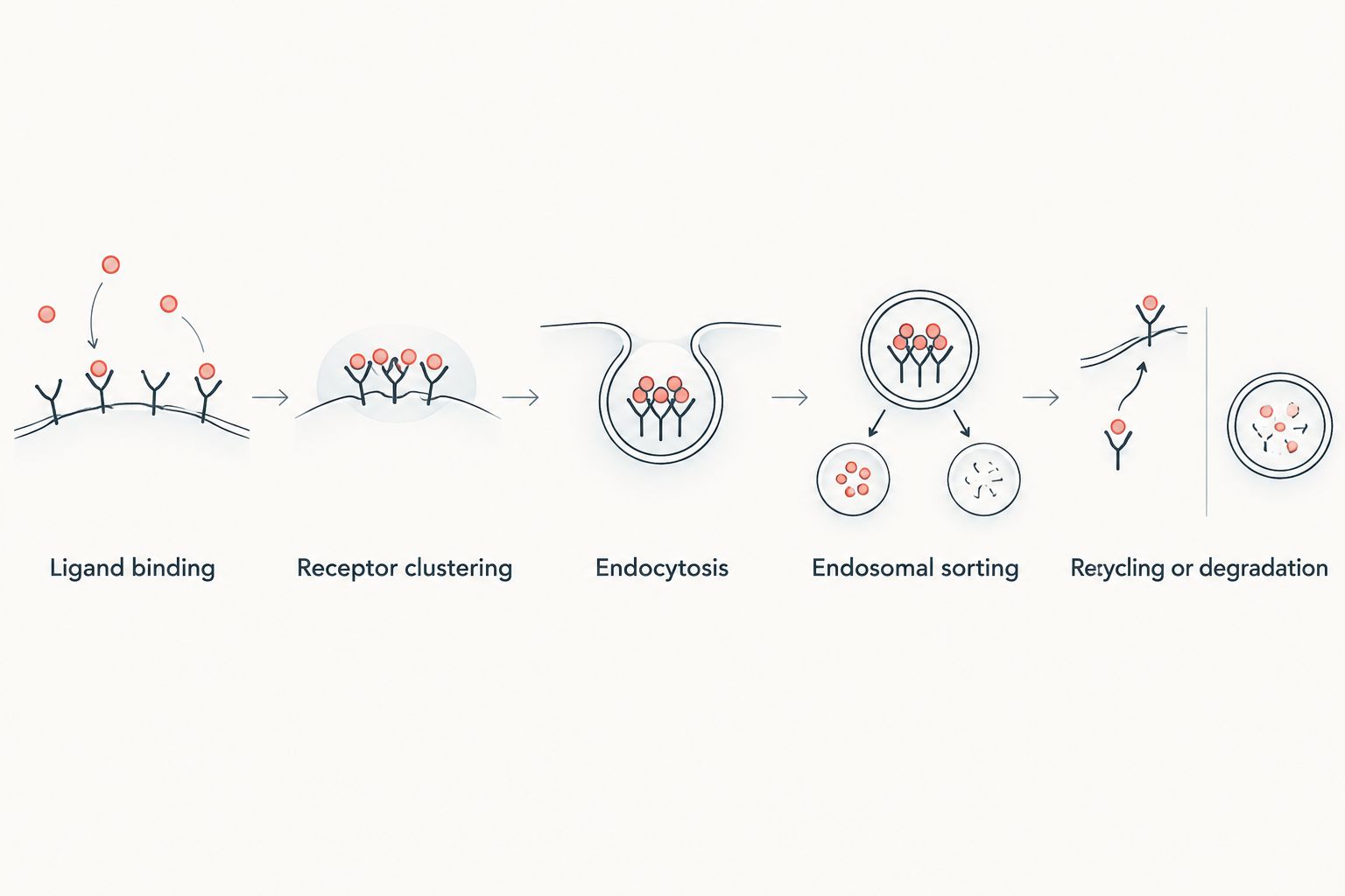
GLP-1 receptor internalization is the agonist-driven movement of the receptor from the cell surface into endosomes, measured by BRET, surface ELISA, or fluorescent imaging to compare peptide analogs and signaling bias. Reproducible curves depend more on peptide purity, consistent reconstitution, and cell line passage control than on the detection platform chosen.
Key points
- GLP-1 receptor internalization assays measure receptor endocytosis after agonist binding, using BRET, ELISA, or imaging readouts.
- Peptide purity and reconstitution consistency drive assay reproducibility more than the detection platform chosen.
- Glp-123 supplies high-purity GLP-1 research peptides with certificates of analysis for internalization and desensitization protocols.
- Biased agonists produce different internalization kinetics than balanced agonists, a confound labs still work around in 2026.
Biotech and academic lab researchers studying GLP-1 receptor internalization measure how fast and how far the receptor moves off the cell surface after agonist exposure, then use that rate to compare peptide analogs, desensitization patterns, and signaling bias. Getting a clean, reproducible internalization curve depends more on assay design and peptide integrity than on any single instrument or software package.
- GLP-1 receptor internalization assays measure receptor endocytosis after agonist binding, using BRET, ELISA, or imaging readouts.
- Peptide purity and reconstitution consistency drive assay reproducibility more than the detection platform chosen.
- Glp-123 supplies high-purity GLP-1 research peptides with certificates of analysis for internalization and desensitization protocols.
- Biased agonists produce different internalization kinetics than balanced agonists, a confound labs still work around in 2026.
- Cell line selection and receptor expression level explain most inter-lab variability reported in internalization data.
Why this matters
GLP-1 receptor internalization is not a side note in receptor pharmacology, it's the mechanism that determines how long a signal from the receptor lasts before the cell resets. When a lab reports that one analog produces a longer cAMP response than another, internalization rate is usually the variable doing the explaining. Understanding GLP-1 receptor structure research gives context for why certain binding pocket differences change how fast the receptor gets pulled into endosomes.
For biotech and academic labs, the stakes are different than for a pharma discovery team running thousands of compounds through automated screens. A university lab or small biotech is usually running a handful of conditions with limited replicates, so every source of noise in the assay shows up directly in the final figure. GLP-1 receptor internalization data is only as reliable as the peptide stock used to generate it.
How to run a GLP-1 receptor internalization study
Select a validated receptor cell line model
Start with a cell line that has documented GLP-1 receptor expression and a known passage-stable phenotype before touching any peptide.
- Confirm receptor copy number is consistent across the passage range you plan to use.
- Check whether the line is native-expressing or a stable overexpression construct, since internalization kinetics differ between the two.
- Match your line choice to prior published work if you want directly comparable results.
- Rule out mycoplasma contamination, which alters membrane trafficking and skews internalization readouts.
Choose a receptor internalization assay format
Decide on a detection method before you order anything, because the format dictates your cell density, timepoints, and reagent needs.
- Surface ELISA for simple endpoint quantification across a dose-response curve.
- BRET or FRET-based assays for real-time kinetic tracking in live cells.
- Fluorescent receptor imaging for visualizing where the receptor goes after it leaves the surface.
- Radioligand binding loss for absolute receptor number, when your lab already handles radioactive materials.
Source a high-purity peptide standard for stimulation
The agonist driving your internalization curve has to be consistent lot to lot, or your kinetics data won't replicate. This is the point where peptide sourcing stops being a logistics question and starts being a data-quality question. Labs building out GLP-1 cell line models for receptor research typically pair the model with a peptide standard that has documented purity and identity confirmation.
- Confirm purity by HPLC and identity by mass spec before the peptide enters your assay.
- Match peptide sequence exactly to the analog you intend to study, including any modifications.
- Request a certificate of analysis for every lot, not just the first order.
- Standardize concentration across replicates instead of eyeballing dilutions from memory.
Glp-123 stocks high-purity peptides for laboratory research protocols with lot documentation intended for exactly this kind of receptor work, which shortcuts the vendor-vetting step many labs spend weeks on.
Standardize reconstitution and handling
Inconsistent reconstitution is one of the most common, least discussed sources of internalization assay variability in 2026 literature.
- Reconstitute at the same volume and diluent every time, and record the exact lot of diluent used.
- Avoid vigorous vortexing, which introduces aggregation that blunts receptor engagement.
- Use the peptide within its documented stable working window after reconstitution.
- Store working stocks at the temperature the manufacturer specifies, not whatever freezer has space.
Control for receptor desensitization and biased agonism confounds
Internalization and desensitization are related but not identical, and conflating them is a common misread in published data. A receptor that desensitizes fast without internalizing much is a different pharmacological story than one that internalizes quickly. Reviewing GLP-1 receptor desensitization alongside your internalization curve helps separate the two mechanisms instead of treating them as one number.
- Run parallel cAMP or arrestin recruitment assays to distinguish desensitization from true internalization.
- Test at least two agonist concentrations to check whether the effect is dose-dependent or saturating.
- Flag any compound with a G-protein versus beta-arrestin recruitment mismatch, since GLP-1 biased agonism changes internalization kinetics independent of overall potency.
- Include a known balanced agonist as a reference point in every run.
Validate results against reference standards
Before you trust a kinetic curve enough to publish or present it, check it against something external.
- Compare your baseline internalization rate to values reported in peer-reviewed literature for the same cell line.
- Re-run a subset of conditions with a fresh peptide lot to confirm the curve holds.
- Document assay temperature, timepoints, and detection antibody lot, since small protocol drifts change absolute numbers.
- Archive raw kinetic traces, not just summary EC50 or half-life values, so the data can be re-analyzed later.
Document a reproducible protocol
A protocol that only works in your own hands isn't finished. Write it so another lab, or your own team in 2026, can reproduce the exact curve.
- List peptide vendor, lot number, and CoA reference alongside every dataset.
- Note cell passage number and seeding density for each experiment.
- Record every buffer and antibody lot used in the detection step.
- Publish the raw timepoints, not just a fitted curve, wherever the venue allows it.
Comparing internalization assay options
| Assay Option | Best For | Key Limitation |
|---|---|---|
| BRET/FRET internalization assay | Real-time kinetics in live cells | Requires a tagged receptor construct and extra cloning step |
| Radioligand binding/surface loss | Absolute receptor number quantification | Radioactive waste handling, lower throughput |
| GFP/fluorescent receptor imaging | Visualizing endosomal trafficking pattern | Needs high-content microscope access |
| Surface ELISA (whole-cell) | Simple screening across dose-response ranges | Endpoint only, no kinetic resolution |
| Biased agonism functional panel | Comparing arrestin versus G-protein pathway internalization | Needs multiple parallel assay reads per compound |
No single format covers every question — a lab chasing kinetics needs BRET or imaging, a lab chasing absolute receptor counts needs radioligand binding, and most labs end up running two formats in parallel to cross-check results.
Common mistakes labs make with GLP-1 receptor internalization studies
- Treating every peptide lot as identical. Purity and HPLC profile can shift lot to lot even from the same vendor, and that shift shows up directly in internalization rate.
- Using reconstituted peptide past its stable window. Aggregated peptide binds inconsistently, which flattens or distorts the internalization curve without an obvious cause.
- Ignoring passage-number drift in the cell line. Receptor expression density changes as cells age in culture, and that alone can explain a curve shift a lab misattributes to the compound.
- Comparing biased agonists to balanced agonists on internalization alone. Without a parallel cAMP or arrestin readout, a biased compound's internalization profile gets misread as simple potency difference.
- Skipping vehicle controls. Constitutive internalization happens without any agonist present, and without a baseline, the agonist-driven signal can't be isolated.
FAQ
What is GLP-1 receptor internalization in research terms?
GLP-1 receptor internalization is the process where the receptor moves from the cell surface into endosomes after agonist binding, ending the surface signal. It’s measured by BRET, ELISA, or fluorescent imaging assays in receptor pharmacology labs.
Is GLP-1 receptor internalization the same as desensitization?
No, desensitization is the loss of receptor responsiveness and internalization is the physical removal of the receptor from the surface. A receptor can desensitize quickly without high internalization, so labs run both assays in parallel to separate the two effects.
Which cell line is best for GLP-1 receptor internalization assays?
The best cell line is one with documented, passage-stable GLP-1 receptor expression matching the assay format you’re running, whether native-expressing or stable overexpression. Consistency across passages matters more than the specific line chosen.
Does peptide purity affect internalization assay results?
Yes, peptide purity and reconstitution consistency are among the largest sources of variability in internalization data. A certificate of analysis for every lot helps rule this out as a confound before blaming the biology.
How does biased agonism change internalization kinetics?
Biased agonists that favor beta-arrestin recruitment over G-protein signaling tend to show different internalization rates than balanced agonists at the same receptor. Running a parallel functional panel distinguishes bias from simple potency differences.
What assay format gives the fastest internalization readout?
BRET or FRET-based assays give real-time kinetic data in live cells, making them the fastest format for tracking internalization as it happens. Surface ELISA is simpler but only gives an endpoint measurement.
Can internalization data be compared across labs?
Comparing across labs is possible only when cell line, passage number, peptide lot documentation, and assay timepoints are reported alongside the data. Without that detail, absolute internalization rates from different labs are not directly comparable.
One last thing
Most internalization curves that fail to replicate trace back to the peptide stock, not the cell biology. A lab chasing a kinetic discrepancy in 2026 should re-check the CoA and reconstitution log before redesigning the assay.
Related guides
Originally published on glp-123.com. This library mirrors the guide for easier reading. Products mentioned are for laboratory research only.
View original


