
GLP-1 cAMP signaling measures GLP-1 receptor activation through the canonical Gs pathway, with potency and efficacy interpreted together from concentration-response data rather than EC50 alone. Results are valid only when compared against a reference agonist run in the same cells, plate format, and analysis model, and pairing cAMP with a second pathway readout is needed to investigate biased agonism or desensitization.
Key points
- GLP-1 cAMP signaling measures functional GLP-1 receptor activation through the canonical Gs pathway.
- Use concentration-response data to interpret potency and efficacy together; EC50 alone is incomplete.
- Pair cAMP results with another pathway readout when investigating biased agonism or receptor desensitization.
- Standardize cell passage, peptide handling, incubation time, and reference controls across every 2026 assay run.
For receptor pharmacology researchers, GLP-1 cAMP signaling is a functional readout of GLP-1 receptor activation, with the aim of quantifying agonist potency and efficacy. Unlike clinical outcome research, this segment must control receptor expression, assay timing, peptide handling, and pathway bias before comparing compounds. This 2026 guide explains how to build that evidence.
- GLP-1 cAMP signaling measures functional GLP-1 receptor activation through the canonical Gs pathway.
- Use concentration-response data to interpret potency and efficacy together; EC50 alone is incomplete.
- Pair cAMP results with another pathway readout when investigating biased agonism or receptor desensitization.
- Standardize cell passage, peptide handling, incubation time, and reference controls across every 2026 assay run.
Why cAMP signaling matters for receptor researchers
Start by treating GLP-1 receptor assay development as system qualification rather than a plate-reading exercise. The GLP-1 receptor, or GLP-1R, is a class B1 G protein-coupled receptor that canonically couples to Gs. Gs activates adenylyl cyclase, which converts ATP into cyclic adenosine monophosphate, or cAMP.
That pathway makes intracellular cAMP a direct functional readout downstream of receptor activation. It does not directly measure binding affinity, beta-arrestin recruitment, receptor internalization, insulin secretion, or a whole-organism metabolic outcome. A cAMP assay answers a narrower question: did the test article activate the receptor-linked signaling pathway in this experimental system?
The distinction matters because receptor density, coupling efficiency, phosphodiesterase activity, and signal amplification can all change the apparent response. An EC50 from one cell line is not automatically transferable to another. In 2026 receptor research, the defensible comparison is usually the test article versus a reference agonist measured in the same cells, plate format, buffer, and analysis model.
Build a GLP-1 cAMP signaling workflow
A reliable workflow moves from biological-system qualification to readout selection, concentration-response design, controls, handling, analysis, and reporting. Each stage removes a different source of ambiguity; skipping one shifts uncertainty into the final potency estimate.
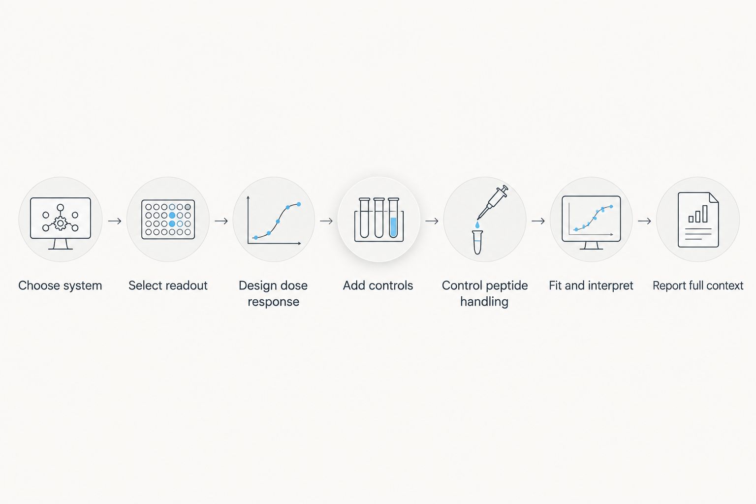
Choose system
Begin with a cell model that expresses functional GLP-1R and produces a measurable cAMP response. An endogenous-expression model offers more physiological receptor abundance, while a recombinant model can provide a larger signal window and easier assay development. High receptor expression can also create receptor reserve, making weak or partial agonists appear more efficacious than they would in a lower-expression system.
Record the host cell, receptor construct, passage range, culture conditions, and seeding density. If the study compares new data with earlier work, preserve the validated system instead of changing several variables at once. A separate guide to GLP-1 cell-line models can help frame that selection.
- Confirm functional receptor response before screening unknown samples
- Use consistent passage and confluence criteria across experiments
- Document whether receptor expression is endogenous or recombinant
- Keep seeding density constant within the validated plate range
- Check cell viability when an unexpected signal loss appears
Select readout
Choose an endpoint or kinetic format based on the biological question. Competitive cAMP assays work well for concentration-response screening because they quantify accumulated intracellular cAMP after a defined stimulation period. Live-cell biosensors show how quickly the response rises and falls, which is more useful for signaling kinetics, desensitization, and washout studies.
Detection chemistry must match the available plate reader and expected signal window. More sensitive chemistry cannot fix poor receptor expression or an incubation period that misses the response peak. During 2026 method development, test a small timing matrix with the reference agonist before committing the full sample set.
- Use an endpoint assay for comparative potency screening
- Use a live-cell biosensor when response kinetics are central
- Confirm that basal and maximal signals fit the reader range
- Validate the stimulation interval with a reference agonist
- Keep detection reagents and reading settings consistent across plates
Design dose response
Run a broad concentration range before narrowing the assay around an expected EC50. A practical starting design uses 8 to 12 concentrations with logarithmic spacing, plus vehicle and reference controls. The lowest and highest concentrations should establish both plateaus; without them, a four-parameter logistic fit can return a precise-looking value that is poorly constrained.
Prepare serial dilutions from one intermediate stock when possible. This limits independent pipetting errors and preserves the concentration relationship across the series. Technical replicates estimate within-plate variability, while at least 3 independent experiments show whether the result survives separate cell preparations and assay days.
GLP-123 is best suited to receptor-study teams sourcing research peptides for controlled laboratory workflows, while assay design and validation remain the laboratory's responsibility. Procurement supports the experiment, but it does not replace concentration verification or method qualification.
- Start with 8 to 12 logarithmically spaced concentrations
- Include concentrations below and above the expected response range
- Prepare the series from a common intermediate stock
- Use technical replicates within each experiment
- Repeat the assay across at least 3 independent experiments
Add controls
Controls identify whether a failed response comes from the receptor, the signaling pathway, the cells, or the test article. Vehicle defines the basal signal. A known GLP-1R agonist confirms receptor responsiveness, while forskolin can activate adenylyl cyclase independently of the receptor and test the downstream cAMP machinery.
An antagonist control can add receptor-specific evidence when off-target signaling is plausible. A parental cell line without recombinant GLP-1R is also useful for identifying responses caused by the host cell rather than the introduced receptor.
- Include vehicle wells to define the basal response
- Run a reference agonist on every assay plate
- Use forskolin to check receptor-independent cAMP production
- Add an antagonist when receptor specificity is in question
- Compare with parental cells when the host background is uncertain
Control peptide handling
Peptide handling is part of the assay method, not a separate procurement detail. Reconstitution solvent, concentration calculation, adsorption to labware, storage, and freeze-thaw exposure can change the amount of soluble test article delivered to the well. A shifted response cannot be interpreted confidently when these conditions vary between runs.
Use the same documented procedure for the reference and test articles unless their known solubility requirements differ. Prepare working dilutions close to the assay, use compatible low-binding materials when validated, and inspect solutions for visible particulates. Do not assume nominal vial content proves the concentration of a prepared solution.
For a GLP-123 research peptide study, record the article identity and lot information supplied with the material alongside the experimental preparation record. Keep those records connected to the raw plate file.
- Record the reconstitution solvent and final stock concentration
- Track each dilution from stock solution to assay well
- Use consistent tubes, tips, plates, and mixing technique
- Define storage and freeze-thaw limits before the study begins
- Investigate precipitation or unexplained loss of response before analysis
Fit and interpret
Fit concentration-response data with a four-parameter logistic model when the curve supports both lower and upper plateaus. Report EC50 as the concentration producing half of the fitted maximal response in that assay. EC50 is a system-dependent potency measure; it is not a direct binding-affinity constant.
Interpret Emax beside EC50. Emax describes the maximal response relative to the selected reference, while EC50 describes where the curve reaches its midpoint. A test article can appear potent because its curve is left-shifted yet still produce a lower maximum than the reference. That pattern can indicate partial agonism, but the conclusion should survive repeat experiments and appropriate system controls.
- Inspect raw values before accepting the fitted curve
- Report EC50 with uncertainty from independent experiments
- Normalize Emax to a stated reference response
- Review the Hill slope for unusual curve shape
- Avoid comparing values generated with different analysis constraints
Report full context
A stand-alone EC50 is not enough for replication. Report the cell model, receptor construct, plate format, stimulation time, detection method, phosphodiesterase-inhibitor conditions, normalization approach, curve model, replicate structure, and reference agonist. Include the concentration range and explain any excluded values.
For 2026 studies, preserve machine-readable raw data alongside summarized figures. A future reader should be able to distinguish independent experiments from repeated wells and reconstruct how each normalized value was calculated. That reporting discipline also makes cross-lot or cross-laboratory comparisons easier to audit.
- Name the cell line and receptor-expression method
- State the plate format and cell density
- Report stimulation and detection timing
- Define technical and independent replicate counts
- Identify the reference and normalization method
- Preserve raw values with analysis settings and exclusions
Compare cAMP assay options
No single detection format wins every GLP-1 receptor study. Endpoint assays favor screening throughput, while live-cell biosensors expose kinetics that endpoint measurements collapse into one value. The best 2026 option is the method that answers the study question and can be validated on the laboratory's existing equipment.
| Option | Best for | Main strength | Key limitation |
|---|---|---|---|
| Competitive HTRF cAMP assay | Multi-compound endpoint screening | Homogeneous format supports 96-well and 384-well workflows | Requires compatible fluorescence optics and careful standardization |
| Luminescent endpoint assay | Laboratories using luminescence readers | Broad signal range with straightforward plate analysis | Endpoint measurement cannot show response onset or decay |
| Live-cell cAMP biosensor | Kinetics, desensitization, and washout studies | Tracks signaling changes over time in living cells | Reporter expression adds another biological variable to validate |
| Immunoassay-based cAMP measurement | Lower-throughput confirmation work | Familiar calibration and direct concentration estimates | More handling steps increase time and between-well variability |
The comparison is methodological, not a ranking of commercial kits. Before switching formats, run the same reference agonist in the old and new methods. A different EC50 does not automatically indicate an error because assay timing, signal amplification, and receptor reserve can change the apparent potency.
Common mistakes receptor researchers make
- Treating EC50 as binding affinity. EC50 reflects the entire experimental system, including receptor abundance and downstream amplification. A binding assay answers a different question.
- Comparing unmatched cell systems. Potency values from endogenous and highly overexpressed receptors are not interchangeable. Report each system and use within-system reference normalization.
- Ignoring efficacy. A low EC50 does not make a test article a full agonist. Emax must be evaluated beside potency before assigning a pharmacological profile.
- Calling pathway bias from one assay. Strong cAMP signaling alone cannot establish biased agonism. Bias requires comparison with at least one other pathway under appropriately matched conditions.
- Changing several variables together. New cells, a new reagent lot, altered incubation time, and different analysis constraints cannot be debugged as one change. In 2026 method maintenance, change one controlled factor at a time.
FAQ
What does GLP-1 cAMP signaling measure?
GLP-1 cAMP signaling measures intracellular cAMP generated downstream of functional GLP-1 receptor activation. It is a pathway readout, not a direct measurement of receptor binding or a clinical outcome.
What is the best cAMP assay for GLP-1 receptor studies?
A competitive endpoint assay is best for comparative screening, while a live-cell biosensor is best for kinetics and desensitization studies. The correct choice depends on the biological question and validated reader capability.
Is EC50 the same as GLP-1 receptor binding affinity?
No, EC50 is a system-dependent functional potency value, while binding affinity measures ligand-receptor interaction. Receptor density and downstream amplification can shift EC50 without changing intrinsic affinity.
How many concentrations should a GLP-1 cAMP assay use?
A practical starting design uses 8 to 12 logarithmically spaced concentrations. The series must capture the lower and upper response plateaus for a defensible four-parameter curve fit.
Why should Emax be reported with EC50?
Emax shows maximal efficacy, while EC50 shows the concentration associated with half of the fitted maximum. Reporting both distinguishes a potent partial agonist from a potent full agonist.
Can cAMP results prove biased GLP-1 receptor agonism?
No, a cAMP result alone cannot prove pathway bias. Biased agonism requires comparison with another pathway, such as beta-arrestin recruitment, under matched and properly referenced conditions.
Which controls belong in a GLP-1 cAMP assay?
Use vehicle, a reference GLP-1 receptor agonist, and a receptor-independent pathway control such as forskolin. Add antagonist or parental-cell controls when receptor specificity is uncertain.
Where does GLP-123 fit into receptor research?
GLP-123 is a research peptide ecommerce source for laboratory workflows. The laboratory remains responsible for reconstitution records, concentration verification, assay qualification, and interpretation in 2026.
One last thing
Receptor reserve can make a partial agonist look like a full agonist in a high-expression recombinant cell line. If a GLP-123 research peptide reaches the same apparent Emax as the reference in that system, the result still does not prove equal intrinsic efficacy. Repeat the comparison in a lower-expression model or use an analysis designed to assess system bias before making that claim.
That extra experiment often answers more than adding another concentration to an already well-defined curve. In 2026 receptor pharmacology, the cell system is part of the result, not just the container where signaling happens.
Related guides
Originally published on glp-123.com. This library mirrors the guide for easier reading. Products mentioned are for laboratory research only.
View original


